Eye Care Services
The team of doctors at Cooper Eye Care in New York City, with offices in Manhattan and Brooklyn serving the surrounding areas of Upper East Side and Dyker Heights, are friendly, professional, and focused on care. As part of a leading eye care practice in New York City, our optometrists offer their experience and talent to their patients in the New York area. The team at Cooper Eye Care includes optometrists, ophthalmologists, and opticians to deliver expert care.
Click on a service below to learn more.
- Comprehensive Eye Exam
- Vision Therapy
- Binocular Vision Problems
- Myopia Control
- Orthokeratology
- Pediatric Eye Care
- Amblyopia
- Dry Eyes
- LASIK Consultation
- Cataract Management
- Nuance Audio Glasses
- Convergence Insufficiency
- Strabismus
- Keratoconus
- Vision Related Learning Disabilities
- Diabetic Eye Disease
- Glaucoma
- Macular Degeneration
Our Specialities

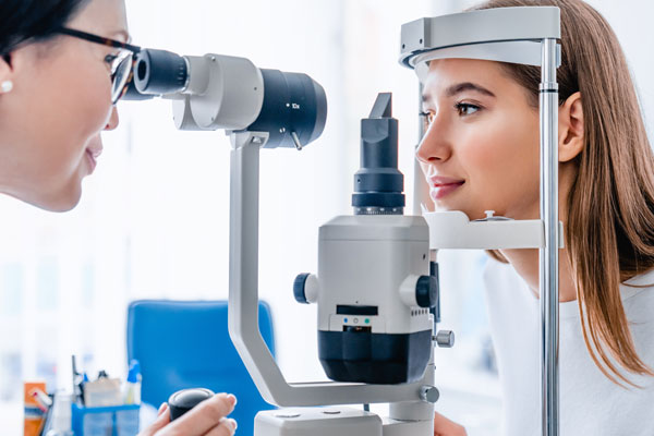
Comprehensive Eye Exam
Comprehensive eye exams are the best way to preserve your healthy vision. The team at Cooper Eye Care strives to make your routine eye exam visits an enjoyable experience. If you’re overdue for a comprehensive eye exam, call the office today to book an appointment.
Learn More
Vision Therapy
While we generally think of glasses, contact lenses, and surgery as the methods of choice to improve vision, there are natural ways to enhance your vision too. At Cooper Eye Care, the skilled optometrists offer vision therapy to improve eye tracking, focusing, teaming and perceptual skills for a lifetime. Weekly in-office therapy supplemented by home therapy can also help treat problems like amblyopia (lazy eye) and strabismus.
Learn More
Binocular Vision Problems
Around 12% of the general population deals with some form of binocular vision problems, including amblyopia, or “lazy eye. We offer comprehensive diagnostic services and treatment options for different types of binocular vision problems. They can also provide relief from associated symptoms, such as dizziness and nausea.
Learn More
Myopia Control
Myopia, or nearsightedness, is the most common refractive error of the eye, affecting upwards of 40% of Americans. At Cooper Eye Care, our team of expert optometrists provides myopia control, a way of combating nearsightedness. Even though there isn’t a cure for myopia, controlling it and curbing its progression provides immeasurable benefits, including relief from eye strain and headaches.
Learn More
Orthokeratology
If you have vision challenges but want to avoid invasive surgical procedures, orthokeratology might be a great fit for you. At Cooper Eye Care, orthokeratology is one of many innovative treatments that can help restore healthy vision. If you’re curious about non-surgical vision correction options, schedule a consultation today.
Learn More
Pediatric Eye Care
We believe in the power of pediatric eye care to prevent eye problems and vision loss in children of all ages. As a parent, ensuring your child stays healthy is a top priority, and routine pediatric eye care is an essential part of that process. Schedule your child’s visit today using the simple online booking tool, or call the office to check appointment availability.
Learn More
Amblyopia
Amblyopia, commonly called lazy eye, is a condition that negatively affects your ability to see properly. Our team of skilled and compassionate eye care specialists offers thorough diagnostic and treatment options for amblyopia. The practice treats both children and adults and works with you to find the best possible solution for your individual set of needs.
Learn More
Dry Eyes
While many health issues with your eyes present few symptoms, dry eye is not one of them. The discomfort and inconvenience of dry eye bring many patients for the latest in dry eye treatment. If over-the-counter lubricating drops are not getting the job done, schedule an appointment to explore more advanced dry eye treatment options.
Learn More
LASIK Consultation
LASIK surgery is the most popular type of refractive surgery in America, and for good reason. The team at Cooper Eye Care have completed hundreds of LASIK procedures, restoring proper vision for men and women. If you’re tired of relying on prescription eyeglasses or contact lenses to get through your daily routines, LASIK might be a great fit for you.
Learn MoreCataract Management
Comprehensive eye exams are the best way to preserve your healthy vision. The team at Cooper Eye Care strives to make your routine eye exam visits an enjoyable experience. If you’re overdue for a comprehensive eye exam, call the office today to book an appointment.
Learn More
Nuance Audio Glasses
Nuance Audio Glasses are not a mere "sound amplifier", but a proper medical device. They are eyewear frames embedded with groundbreaking technology which seamlessly integrate an air conduction hearing aid intended to amplify sound.
Learn More
Convergence Insufficiency
Convergence insufficiency, the inability of your eyes to work together and focus on nearby objects, is a leading cause of eye strain, blurred vision, and double vision. The skilled optometrists at Cooper Eye Care diagnose and treat this troublesome condition effectively with solutions like prescription lenses and comprehensive vision therapy. If activities such as reading strain your eyes, call or schedule an appointment online today to find out if you have convergence insufficiency.
Learn More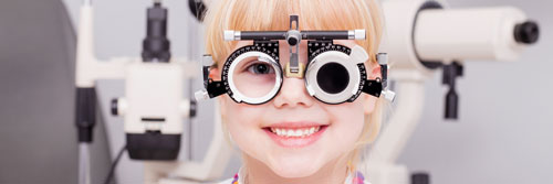
Strabismus
Approximately 4% of children in the United States suffer from strabismus, a visual problem marked by eyes pointing in different directions. At Cooper Eye Care, we can treat this condition for patients of all ages. Strabismus can be diagnosed during an eye exam and there are several treatment options available, including prescription lenses and corrective surgery.
Learn More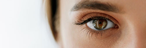
Keratoconus
Keratoconus is defined as progressive thinning and protrusion of the cornea. The cornea is the front surface of the eyeball. It is typically round, relatively uniform in thickness, and clear. In a normal eye, light passes through the cornea and forms a clear image on the retina, the back of the eye. The retina then transfers the image information to the brain. In an eye that has keratoconus, the cornea is no longer round but protrudes forward and becomes more cone-like. When light passes through the cornea of a keratoconic eye, it becomes distorted so that it no longer creates a clear image on the retina. This results in the symptoms of keratoconus.
Learn More
Vision Related Learning Disabilities
Vision plays an integral role in learning, particularly for schoolchildren. While schools aren’t always equipped to diagnose vision-related learning disabilities, the skilled optometrists at Cooper Eye Care can diagnose these issues through comprehensive eye exams. The staff routinely helps address vision-related learning disabilities, one of the most prevalent childhood conditions, with treatment options like corrective lenses, vision therapy, and more.
Learn More
Diabetic Eye Disease
Diabetes can lead to a range of health issues, including several that affect your eyes. Our team of trained eye care professionals provide comprehensive treatment for different forms of diabetic eye disease, such as cataracts and glaucoma. Without medical intervention, diabetic eye disease can cause severe vision loss or even blindness.
Learn More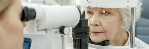
Glaucoma
An estimated 3 million Americans have glaucoma, and only half of them are aware they even have the condition. At Cooper Eye Care, men and women have access to a team of highly skilled eye health specialists that can assist in diagnosing and treating glaucoma. If you’re overdue for an eye examination, schedule an appointment today to preserve your healthy vision.
Learn More
Macular Degeneration
According to the National Eye Institute, age-related macular degeneration is one of the most significant causes of vision loss in the United States and is projected to affect more than 5 million Americans by 2050. The prevalence of this serious eye health concern is why the team at Cooper Eye Care offers comprehensive diagnostic and treatment of macular degeneration at their offices in Brooklyn and Manhattan.
Learn More